



You've got the plasmid prep on ice, the cells look healthy enough, and the reporter assay is waiting. The annoying part is that last week's setup worked, this week's didn't, and nothing obvious changed except the part that always seems minor until it isn't. That's usually where transfection of plasmid becomes less about “adding DNA to cells” and more about understanding the barrier, the cargo, and the cell type in front of you.
A plasmid is a circular DNA vehicle that carries a gene or other sequence into a eukaryotic cell so the cell can express it transiently or, in some cases, integrate it stably. The core idea is simple, but the outcome depends on how the DNA gets across the membrane, what state the cell is in, and whether the DNA itself is clean enough to do its job. The method matters because the same delivery approach that works in HEK293T cells can fall apart in primary, suspension, hematopoietic, or stem cells.
Table of Contents
- What is the difference between transfection and transformation
- Should I choose stable or transient transfection
- Are viral vectors ever justified for a one-off experiment
- How do I know if I should switch from chemical to electroporation
- Where does AI or connected lab data fit into transfection optimization
What Plasmid Transfection Actually Means
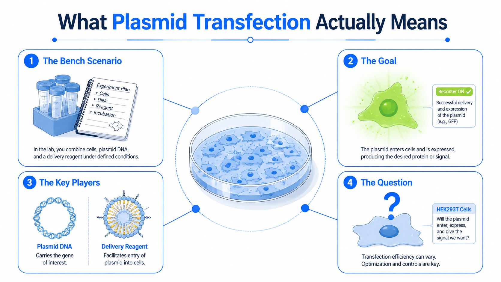
A plasmid is the payload, but the cell membrane is the primary problem. In plasmid transfection, you deliberately introduce that circular DNA into a eukaryotic cell so the cell can read it, express it, and sometimes keep it long term if the DNA integrates. The method is the bridge between a tube of purified DNA and a living cell that can effectively use it.
Plasmid transfection is not transformation
People often mix up transfection and transformation because both involve foreign DNA. In bacteria, transformation means getting DNA into a microbial host, usually for cloning or propagation. In mammalian or other eukaryotic cells, transfection is the term used for introducing nucleic acid into cells that are much more sensitive to how that DNA arrives.
That distinction matters because the cell environments are different. Bacteria can be handled one way, while mammalian cells need delivery systems that avoid excessive damage. The plasmid itself doesn't change the logic, but the membrane, intracellular trafficking, and toxicity limits absolutely do.
What the plasmid is doing inside the cell
Once inside, a plasmid usually serves as a temporary instruction set. It can drive gene expression, produce a reporter signal, or support longer-term selection if the construct is designed for stable integration. In practical terms, you're not “feeding” the cell DNA, you're trying to get the cell to make a protein, show a phenotype, or provide a genetic edit template.
Practical rule: if the cells look fine but the readout is weak, the issue may be delivery, DNA quality, or the plasmid design itself, not just the reagent.
That's why delivery method matters. The plasmid is the same basic cargo, but the route in changes the chance of uptake, the chance of survival, and the kind of cells you can work with. A good protocol is less about forcing every cell into the same routine and more about matching the method to the biology in front of you.
How Plasmid Transfection Came to Be
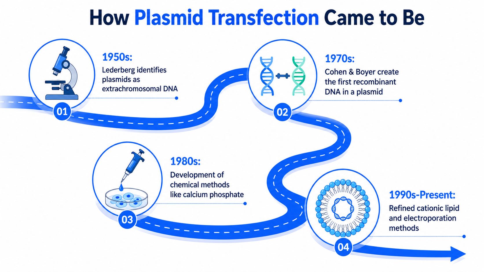
A new lab member often meets plasmid transfection as a routine protocol, but the method grew out of a long sequence of ideas about how DNA moves, survives, and functions inside cells. The early story began before transfection was a standard word in bench language. In the early 1950s, Joshua Lederberg's laboratory helped establish plasmids as autonomous, extrachromosomal DNA, and Lederberg proposed the term “plasmid” in 1952. That concept mattered because it gave researchers a way to think about circular DNA as something that could exist independently of the main chromosome, which later made plasmid DNA a standard laboratory vector for gene delivery into cells. Historical overview of plasmids and recombinant DNA
From bacterial genetics to mammalian delivery
The next step came between 1972 and 1974, when Stanley Cohen and Herbert Boyer used plasmids to build recombinant DNA molecules by cutting a plasmid, inserting foreign DNA, resealing it, and putting it back into bacteria. That work became the experimental basis of recombinant DNA technology and modern genetic engineering. By the 1970s, plasmids were already being used to insert genes into bacteria for production of therapeutic proteins such as human insulin.
Mammalian delivery followed a different path. The first report of nucleic acid uptake by mammalian cells using DEAE-dextran appeared in 1962, then calcium phosphate co-precipitation enabled the first successful gene transfer into mammalian cells in 1973, with stable integration shown in 1977. Electroporation arrived in 1983, using electrical pulses to increase membrane permeability, and later the field expanded beyond DNA to include mRNA and proteins in the 2010s, with importance in CAR-T manufacturing and personalized therapies by 2012. Transfection timeline of key events
These milestones explain why the same plasmid can behave differently across cell types. A bacterial host tolerates one set of entry conditions, while a mammalian cell cares about membrane stress, endosomal escape, and how much injury it can recover from. The method is never just about the DNA, it is about matching the delivery route to the biology of the cell in front of you.
Why that history still matters at the bench
The common thread is a circular DNA cargo crossing a membrane barrier without tearing the cell apart. Each method changed the physical route, but the goal stayed the same, get nucleic acid into a cell and keep the cell functional afterward. That is why modern plasmid transfection still draws from both bacterial genetics and delivery engineering.
For a new experiment, the practical lesson is straightforward. The best method is the one that fits the cell type, the readout, and the amount of stress the cells can tolerate. Easy adherent lines, difficult primary cells, suspension cultures, and stem cells all ask for different trade-offs, so the history of plasmid transfection is really a history of learning how to work with that variation instead of pretending it does not exist.
Major Delivery Methods Side by Side
Different delivery methods solve different bottlenecks. Some are built for speed, some for fragile cells, and some for scale. If you treat all of them like interchangeable bottles on the same shelf, the protocol will punish you later.
Mechanism first, then fit
Chemical methods, like calcium phosphate and lipofection, package DNA into complexes that cells can take up more easily. Lipid systems act like molecular envelopes, helping DNA cross the membrane and escape endosomes. Electroporation uses a short electrical pulse to open transient pores, which can help when chemistry alone is too gentle. Microinjection puts material directly into a cell, while biolistic delivery uses physical force to shoot particles into tissue or cells. Viral vectors are a separate benchmark, because they're built for efficient genetic delivery, but they come with a very different regulatory and design burden.
A practical comparison lens
| Method | Mechanism | Best-fit cells | Typical efficiency | Main limitation |
|---|---|---|---|---|
| Calcium phosphate | DNA precipitate enters cells by uptake | Many adherent lines | Variable | Sensitive to cell state and handling |
| Lipofection | Lipid-DNA complexes enter by membrane interaction | Common adherent mammalian cells | Often strong in easy lines | Toxicity and cost can rise in sensitive cells |
| Electroporation | Electrical pulse opens membrane pores | Suspension, primary, and harder cells | Often useful when chemistry fails | Can injure cells if conditions are off |
| Viral vectors | Virus-derived particle delivers cargo | Hard-to-deliver cells and some specialized workflows | Very high in many contexts | Complex design, oversight, and payload limits |
| Microinjection | Direct physical injection into cells | Large cells or single-cell studies | Very targeted | Low throughput |
| Biolistic delivery | DNA-coated particles are shot into cells | Tissues and some plant systems | Context dependent | Physical damage and uneven delivery |
For a routine reporter assay in an easy adherent line, lipid-based delivery often makes sense. For primary or suspension cells, electroporation may be the cleaner starting point because membrane access is the bigger issue than uptake chemistry. Viral systems remain useful when the biology demands them, but they're not the default answer for a one-off plasmid readout.
Decision rule: choose the method by cell type first, then by readout, then by how much stress the cells can tolerate.
A Stepwise Workflow That Works in Practice
Good plasmid transfection starts before the DNA and reagent ever meet. The day before, check whether the cells are healthy, attached well, and at a density that gives them room to respond. For many adherent workflows, the common starting point is around the middle of the growth phase rather than overgrown or sparse cultures, because both extremes can hurt uptake and expression.
Build the run around clean inputs
Plasmid quality matters more than many new users expect. Check the DNA concentration, look at the A260/280 ratio, and pay attention to endotoxin because contamination can sharply reduce efficiency, especially in primary cells, suspension cells, and hematopoietic cells. For transient expression, supercoiled plasmid DNA is generally preferred, while linear DNA can integrate more efficiently for stable transfection, even though uptake is usually lower. QIAGEN transfection guidance for DNA
When you form complexes, use the medium recommended for the reagent, usually a reduced-serum formulation. Add the complexes dropwise so they spread evenly across the well instead of landing as a local overdose. One published HEK293T protocol uses 1 µg plasmid DNA with 2 µL P3000 and 3 µL Lipofectamine 3000 per 3.5 cm dish, then leaves the cells for 6 h before switching to fresh medium and collects expression at 24 to 36 h. HEK293T Lipofectamine 3000 protocol
What to record every time
- Cell state at plating, including health, density, and morphology.
- Plasmid source and purity, including endotoxin status and prep type.
- Exact DNA-to-reagent ratio, because that ratio usually needs re-optimization.
- Incubation time before medium change, since overexposure often increases stress.
- Readout window, because different assays peak at different times.
The details matter because a protocol that works once is not yet a method. It becomes a method when you can reproduce the outcome and explain what changed when it didn't.
For users who prefer a simple bench refresher while setting up reagents, Herbilabs Labware's usage guide is a useful example of how clear handling instructions can reduce avoidable variation in day-to-day workflows.
Optimizing for Difficult and Sensitive Cells
A protocol that performs well in HEK293T cells can fall apart in primary cells, suspension cultures, hematopoietic lineages, or stem cells. Those systems are less forgiving, so small shifts in DNA quality, cell state, or delivery chemistry can decide whether the experiment works at all. The usual mistake is to keep the same setup and hope the new cell type behaves like the old one.
The better approach is to start by matching the method to the cell, then tune the inputs around that match. A gentle reagent may suit one primary population, while another needs a different delivery route altogether. The workflow is not about forcing every cell into the same mold. It is about finding the narrow conditions that let the cargo enter without pushing the cells past their limit.
Start with DNA purity and topology
Endotoxin contamination can sharply reduce performance in sensitive cells, so a clean prep is the starting condition, not a nice extra. The topology of the plasmid matters too. Supercoiled DNA is usually the better choice for transient expression because it supports expression well, while linear DNA is often better when stable integration is the goal.
That distinction is easy to miss if you mostly work in forgiving lines. In fragile systems, the same plasmid can look “bad” because the cell is stressed, not because the construct is flawed. Remove obvious contamination first, then decide whether the experiment needs short-lived expression or stable integration.
Re-optimize instead of borrowing ratios
The DNA-to-reagent ratio is not portable across every cell line and every plasmid. Guidance from QIAGEN notes that the optimal ratio needs to be re-optimized for each new cell line and plasmid combination, and difficult cells often need more care than the vendor example you may have used before. A small test matrix is usually better than copying a ratio from a forgiving system and expecting it to hold.
Test a few ratios, keep the rest of the system fixed, and change only one variable at a time. That is how you learn whether the problem sits in the DNA, the reagent, or the cell state.
A practical decision tree is straightforward. Start with endotoxin-free plasmid, choose the delivery method that fits the cell type, screen several ratios, and read out both expression and toxicity. If viability drops before expression rises, the delivery chemistry is too aggressive for that cell state. If expression is weak but the cells still look healthy, the barrier is probably uptake rather than toxicity.
Controls, Readouts, and Biosafety Essentials
A transfection result is only useful if you can tell what caused it. That's why the control set matters as much as the reagent choice. Without it, a weak signal could mean poor delivery, a dead promoter, a toxic construct, or a readout problem.
The minimum control set
- Untransfected cells, to show baseline morphology and assay background.
- Reagent-only control, to isolate reagent toxicity.
- Empty-vector control, to separate vector backbone effects from insert effects.
- Positive reporter control, such as GFP or luciferase, to show the system can work.
- Negative silencing or loss-of-function control, when the assay depends on repression or editing.
Each one blocks a different failure mode. Untransfected cells tell you the starting condition. Reagent-only wells tell you whether the chemistry is hurting the cells. Empty vector shows whether the backbone itself changes the outcome. The positive control confirms the cell line can support the assay.
Match the readout to the question
Use fluorescence when you need cell-level distribution or easy visual screening. Use luminescence when sensitivity and dynamic range matter more than imaging. Use qPCR when transcript abundance is the question, Western blot when protein level matters, and a functional assay when you care about what the gene product does. A reporter signal is not always enough, especially if you need to know whether expression is translating into phenotype.
Biosafety still applies
Plasmids are routine tools, but routine doesn't mean risk-free. Check the plasmid's risk group, look closely at the insert, and think about viral homology, toxin genes, and selectable markers before you start. Waste handling and institutional oversight still matter, especially when the construct could change cell behavior in ways that are easy to underestimate at the bench.
Troubleshooting and Metrics for Success
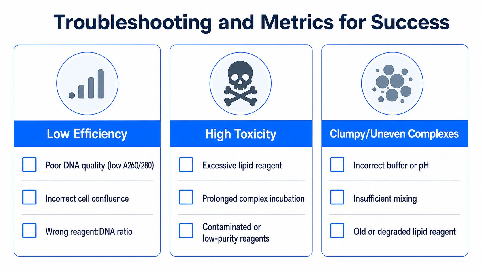
The notebook usually fills up with the same three problems. Low efficiency, high toxicity, and clumpy or uneven complexes account for most of the frustration because each one can look like the others if you don't diagnose them carefully. The fix starts by separating the symptom from the cause.
Low efficiency
If expression is weak, look first at poor DNA quality, incorrect cell confluence, and wrong reagent-to-DNA ratio. The first move is usually to verify the prep, then check whether the cells were too dense or too sparse, then retest the ratio with the same construct. If you change everything at once, you won't know what helped.
High toxicity
If the cells are stressed or detaching, the usual suspects are excessive lipid reagent, prolonged complex incubation, or contaminated or low-purity reagents. Cut back the reagent amount before you abandon the method, and shorten the exposure if the protocol leaves complexes on too long. In sensitive lines, a healthy-looking transfection mix can still be too harsh for the cells.
Clumpy or uneven complexes
If the complexes look inconsistent, check the buffer or pH, whether the mix was insufficiently mixed, and whether the lipid reagent is old or degraded. Uneven complexes often mean the chemistry wasn't allowed to assemble properly, or the reagent lost performance during storage. The solution is usually boring and effective, fresh reagents, correct buffer, and gentle but thorough mixing.
Success is a pattern, not a single readout. You want consistent expression, acceptable viability, and similar performance across replicates, not just one bright well that looks impressive on the microscope.
A strong run leaves you with data you can compare. Log the cell type, plasmid prep, ratio, incubation time, expression window, and toxicity outcome, then adjust only one or two variables on the next pass. That loop, log, compare, adjust, repeat, is what turns transfection from guesswork into a controlled assay.
Common Questions About Plasmid Transfection
What is the difference between transfection and transformation
Transformation is the term usually used for introducing DNA into bacteria. Transfection is the term used for introducing nucleic acids into eukaryotic cells, such as mammalian cells. If you're working with HEK293T, primary cells, or stem cells, you're talking about transfection.
Should I choose stable or transient transfection
Choose transient transfection when you want fast expression, a reporter assay, or a short-term perturbation. Choose stable transfection when you need long-term expression or a cell line you can return to repeatedly. The earlier section on DNA topology matters here, because supercoiled DNA usually helps transient expression, while linear DNA is often better when integration is the goal.
Are viral vectors ever justified for a one-off experiment
Yes, but only when the biology demands it. If the target cells are hard to transfect, or if the experimental question needs very efficient delivery, viral vectors can be justified as a benchmark or a tool. For a simple plasmid readout in a permissive cell line, they're usually more complexity than you need.
How do I know if I should switch from chemical to electroporation
If the cell type is sensitive, suspension-based, or otherwise resistant to chemical uptake, electroporation often deserves a look. It changes membrane permeability directly, so it can solve a problem that lipids or calcium-based methods can't. The trade-off is stress, so viability has to stay part of the decision.
Where does AI or connected lab data fit into transfection optimization
Teams are starting to use connected experimental data to compare transfection conditions across cell lines, plasmids, and reagent systems more systematically. That matters because optimization is rarely about a single magic condition, it's about learning which variables move the outcome in your lab. If your records are fragmented, it's harder to see those patterns, and harder to reuse them.
If you're setting up plasmid transfection as a repeatable platform instead of a one-off experiment, visit Polymerize. It helps teams unify experimental data, connect outcomes across conditions, and use explainable models to plan the next best experiment instead of starting from scratch every time.
Put your R&D data to work
See how materials teams use Polymerize to plan fewer experiments and reach target properties faster.